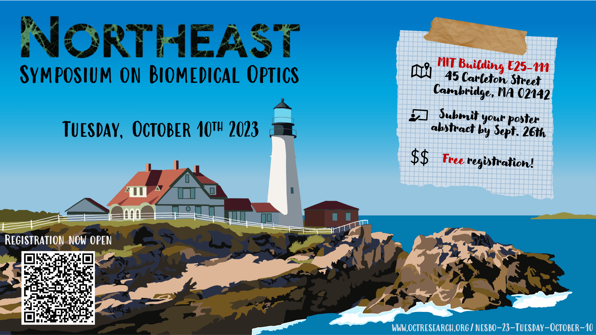
NESBO ’23 Tuesday October 10
About:
NESBO is an annual event aiming to bring together junior researchers from across the greater New England area to stimulate scientific discussion and promote collaboration within the local Biomedical Optics community. It is a great opportunity to interact with other young researchers in a relaxed atmosphere. We hope to see you there!
Registration is now closed.
Please reach out to one of the organizers for questions.
Schedule
Time | Event | Speaker | Affiliation |
|---|---|---|---|
| 08:30 | Coffee + Badge Distribution | ||
| 09:00 | Introduction | Benjamin J. Vakoc | MGH-Wellman |
Session 1: Translational Biomedical Optics | |||
| 09:15 | Mapping of collagen organization in pulmonary fibrosis in vivo using optic axis-resolved polarization-sensitive endobronchial optical coherence tomography | Jaeyul Lee | MGH-Wellman |
| 09:40 | Perfluorocarbon nanodroplets for photoacoustic image-guided oxygen-enhanced photodynamic therapy of hypoxic tumors | Marvin Xavierselvan | Tufts University |
| 10:05 | Break | ||
Session 2: Measuring Small Phase Fluctuations | |||
| 10:30 | Characterizing 3D micromechanical heterogeneity in the extracellular matrix with photonic force optical coherence elastography | Nichaluk Leartprapun | MGH-Wellman |
| 10:55 | Optical Coatings for Gravitational-Wave Detectors | Nicholas Demos | MIT LIGO Laboratory |
| 11:20 | Panel: Academia versus Industry |
|
|
| 12:20 | Lunch/Discussion | ||
Session 3: Advances in Neuroimaging – I | |||
| 13:20 | Deep brain catheter-based polarization sensitive optical coherence tomography for guiding stereotactic neurosurgery | Shadi Masoumi | Université Laval |
| 13:45 | A miniaturized fluorescence computational imaging platform for extended depth of field neural imaging | Joseph Greene | Boston University |
| 14:10 | Coffee Break | ||
Session 4: Advances in Neuroimaging – II | |||
| 14:20 | HOLiS: Human brain optimized light sheet microscopy for high-throughput cell-type atlasing of whole human brains | Malte Johannes Casper | Columbia University |
| 14:45 | Functional development of motor and sensory circuits for cardiac control | Luis Hernandez-Nunez | Harvard University |
| 15:10 | Poster Session | ||
| 16:40 | Concluding Remarks and Awards | ||
| 17:10 | Social Hour @ Cambridge Brewing Co.(17:00 – 19:00, click for location) | ||
Invited Talks
Translational Biomedical Optics
Mapping of collagen organization in pulmonary fibrosis in vivo using optic axis-resolved polarization-sensitive endobronchial optical coherence tomography
Jaeyul Lee, MGH-Wellman
Click to read abstract
Fibrotic interstitial lung diseases (fILD), such as idiopathic pulmonary fibrosis (IPF), are characterized by abnormal collagen deposition. Assessment of collagen fiber orientation could serve as a biomarker for prognosis and therapeutic responsiveness. Endobronchial optical coherence tomography (EB-OCT) is a bronchoscope-compatible imaging modality that provides volumetric visualization of large tissue volume with microscopic resolution. Polarization-sensitive EB-OCT (PS-EB-OCT) additionally detects endogenous birefringence from organized tissue such as collagen. We previously evaluated PS-EB-OCT for in-vivo microscopic visualization of birefringent fibrosis in fILDs. Here, we further investigate depth-resolved optic-axis (OA) orientation mapping to assess localized collagen fiber orientation in fILDs in-vivo.
Click to read speaker bio
Jaeyul Lee is a postdoctoral research fellow in Dr. Lida Hariri’s group at Division of Pulmonary and Critical Care Medicine, Massachusetts General Hospital and Harvard Medical School. He received the Ph.D. degree in School of Electronic and Electrical Engineering from Kyungpook National University, Daegu, South Korea, in February 2021. Then, he started his first postdoc position at Department of Bioengineering, University of California, Los Angeles, for a year. His research interests are in the development of functional optical imaging techniques, including optical coherence tomography, photoacoustic microscopy,spectroscopy, and their biomedical pulmonary applications.

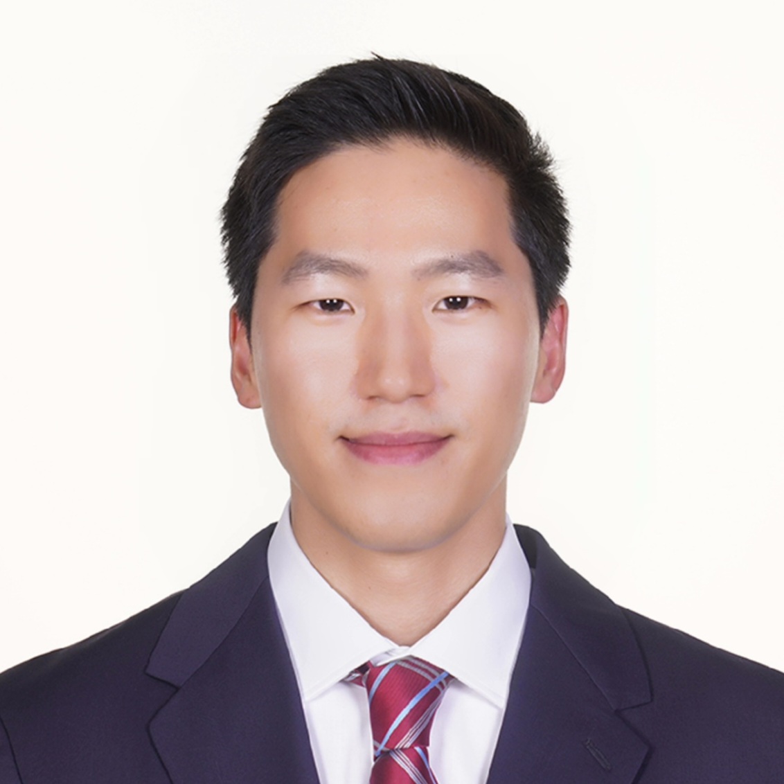
Perfluorocarbon nanodroplets for photoacoustic image-guided oxygen-enhanced photodynamic therapy of hypoxic tumors
Marvin Xavierselvan , Tufts University
Click to read abstract
Solid tumors face the challenge of hypoxia, stemming from an imbalance between oxygen demands and supply. Hypoxia leads to resistance to conventional cancer therapies, such as radiation and chemotherapy, including photodynamic therapy (PDT) which relies on oxygen radicals. To address this challenge, we developed perfluorocarbon nanodroplets for co-delivering oxygen and a photosensitizer. We validated oxygen release and enhanced tumor oxygenation in both in vitro and in vivo models. Histological analysis confirmed reduced hypoxic regions in nanodroplets-treated tumors while PDT using the same resulted in superior efficacy compared to a liposomal formulation. Overall, oxygen-loaded nanodroplets guided by photoacoustic imaging offer a promising approach for treatment of hypoxic tumors.
Click to read speaker bio
Marvin Xavierselvan is a 4th year Ph.D. student at Tufts university’s integrated Biofunctional Imaging and Therapeutics (iBIT) lab. He has experience in designing biomedical instrumentation, photoacoustic imaging, and photodynamic therapy (PDT). His research focuses on photoacoustic imaging and photodynamic therapy to combat resistant tumors. His scientific interests span photoacoustic imaging, image-guided PDT in head and neck, and pancreatic cancer. His Ph.D. thesis aims to enhance PDT efficacy through nanoplatforms, addressing tumor hypoxia and enabling combination therapies with oxygen delivery.
Measuring Small Phase Fluctuations
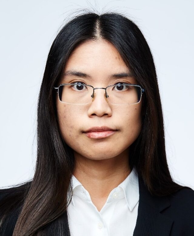
Characterizing 3D micromechanical heterogeneity in the extracellular matrix with photonic force optical coherence elastography
Nichaluk Leartprapun, MGH-Wellman
Click to read abstract
We demonstrate a unique application of an unconventional mode of optical manipulation, radiation pressure from a low-numerical aperture beam, to address the need in the field of mechanobiology for quantitative imaging of extracellular matrix (ECM) mechanics at cellular resolution. Photonic force optical coherence elastography (PF-OCE) utilizes harmonically-modulated radiation pressure to locally “push” on individual microbeads embedded in the ECM. Amount to a few piconewtons, the radiation pressure force induces sub-nanometer bead displacements, demanding state-of-the-art interferometric detection provided by phase-sensitive optical coherence tomography.We validate PF-OCE against shear rheometry in polyacrylamide hydrogels, then, demonstrate micromechanical imaging in 3D ECM-derived hydrogels.
Click to read speaker bio
Nichaluk Leartprapun is a postdoctoral research fellow in the laboratory of Dr. Seemantini Nadkarni at the Wellman Center for Photomedicine, Massachusetts General Hospital. Her current research focuses on the development and applications of laser Speckle rHEologicAl micRoscopy (SHEAR) technology to characterize micromechanical properties of tissues, biofluids, and engineered biomaterials. Prior to joining Wellman, she completed her PhD under the supervision of Dr. Steven Adie at Cornell University, where she developed imaging technologies based on optical coherence tomography and elastography for micromechanical and structural imaging of biological systems.
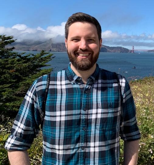
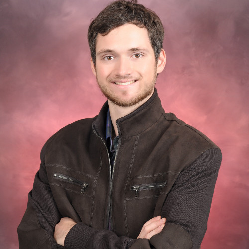
Optical Coatings for Gravitational-Wave Detectors
Nicholas Demos, MIT LIGO Laboratory
Click to read abstract
With the direct detection of gravitational waves from coalescing pairs of binary black holes and neutron stars, the Laser Interferometer Gravitational-Wave Observatory (LIGO) has successfully measured strains smaller than 1 part in 10^21. Although this incredible achievement makes LIGO one of the most sensitive experiments ever created, there is still room to improve. One of the strongest limits placed on LIGO’s sensitivity comes from the optical coatings used to make the mirrors of the interferometer. In this talk, I will describe gravitational waves and LIGO, optical coatings and how to test them, and some ideas for future improvements.
Click to read speaker bio
Nicholas is a physics graduate student at MIT, where he is engaged in research at the Laser Interferometer Gravitational-Wave Observatory (LIGO) Laboratory. His work on optical coatings aims to address fundamental limitations to LIGO’s sensitivity. During his time at MIT, he has been honored with the Barish-Weiss Fellowship and the MathWorks Science Fellowship. A Southern California native, Nicholas completed his undergraduate physics degree at California State University, Fullerton, where he worked on simulations of merging binary black holes as part of the Simulating eXtreme Spacetimes (SXS) collaboration.
Advances in Neuroimaging – I
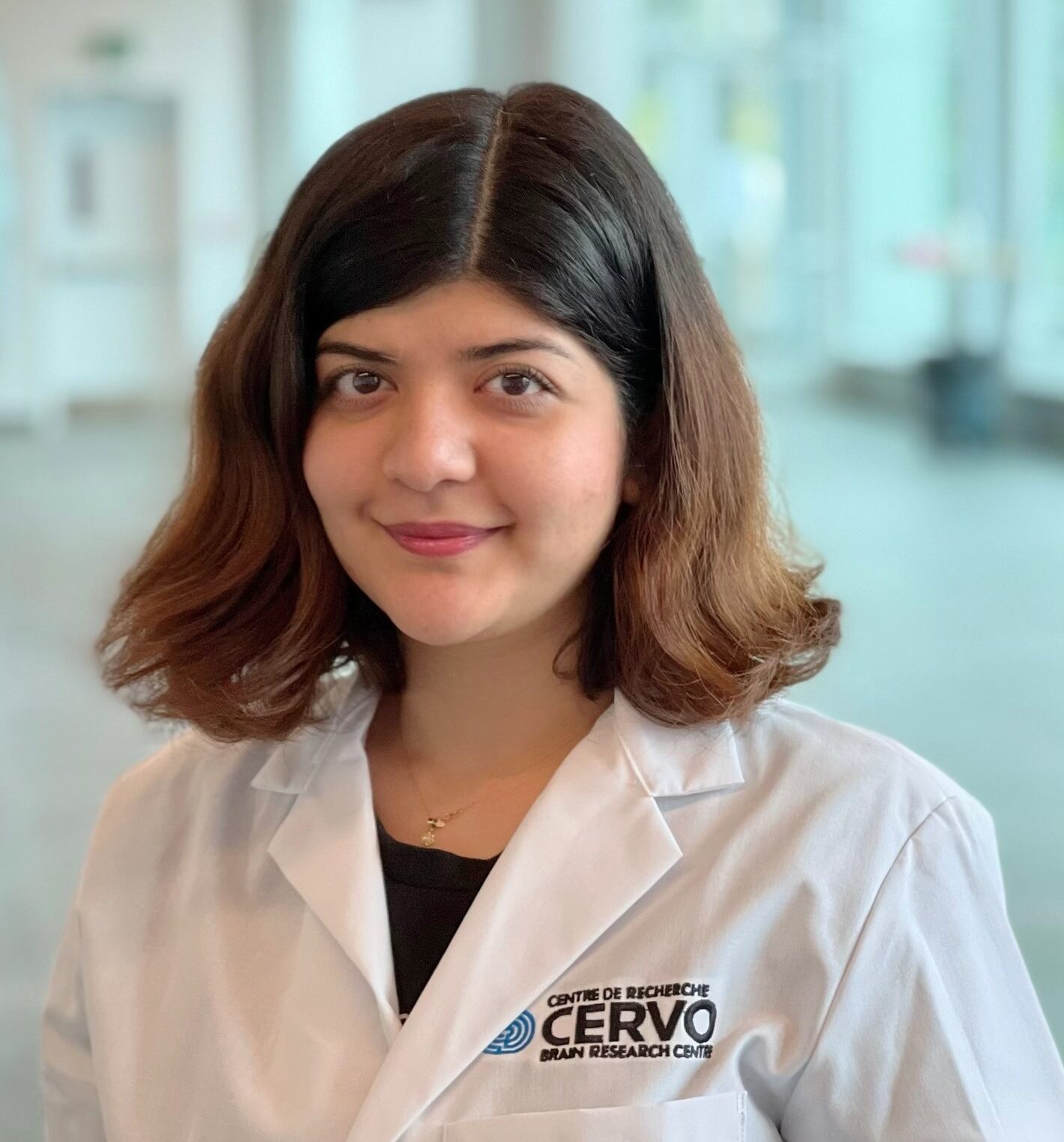
Deep brain catheter-based polarization sensitive optical coherence tomography for guiding stereotactic neurosurgery
Shadi Masoumi, Université Laval
Click to read abstract
Reconstructed depth-resolved optic axis orientation obtained by catheter based PSOCT in a tissue volume informs on the orientation of the white matter fiber bundles in the brain, owing to the birefringence of myelinated axons. The physical organization of white matter also leads to anisotropic diffusion of water molecules, which is the basis of dMRI for non-invasive imaging of the three-dimensional orientation of white matter fiber bundles. Having access to fiber orientation in both imaging modalities, we are trying to map the depth-resolved birefringence and optic axis orientation to the larger scale dMRI as well as an atlas of brain anatomy.
Click to read speaker bio
Shadi is a Fourth year Ph.D. student in Biophotonics at CERVO brain research center, Quebec City, Canada. In collaboration with Prof. Martin Villiger at Wellman Center for Photomedicine,she is using Polarization Sensitive Optical Coherence Tomography (PS-OCT) as a tool for neurosurgical guidance.To do so, she is working on both PS-OCT processing development and mapping PS-OCT to diffusion MRI of the brain. She obtained the B.Sc. in Physics and the M.Sc. in Photonics where she worked on diffuse reflectance spectroscopy of skin.
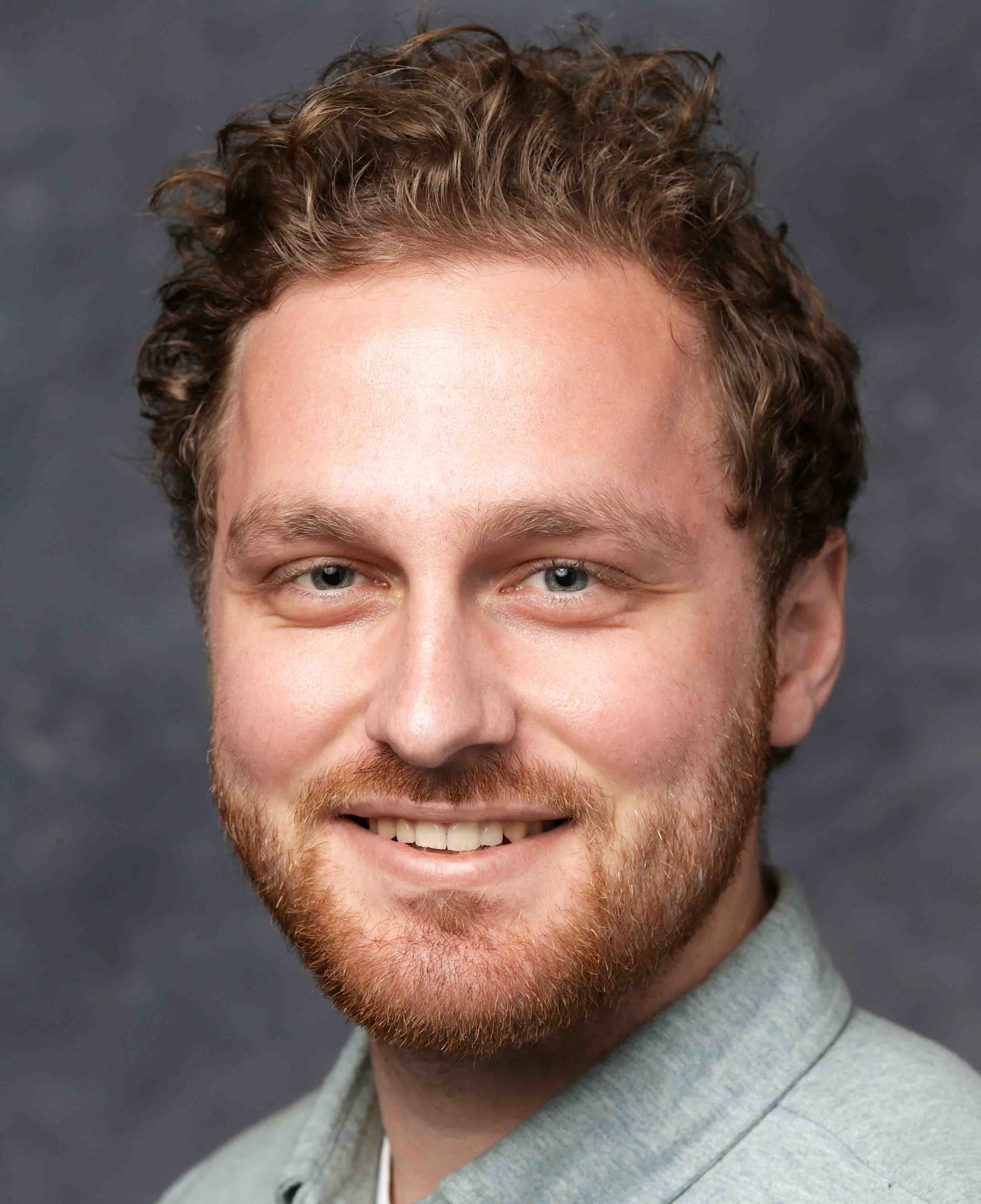
A miniaturized fluorescence computational imaging platform for extended depth of field neural imaging
Joseph Greene, Boston University
Click to read abstract
Miniaturized head-mounted fluorescence imaging platforms, known as miniscopes, push fundamental neuroscience by monitoring real time neural activity in freely behaving animals. However, these platforms inherently exhibit a shallow depth of field due to the use of highly aberrated off-the-shelf optics, 1-photon excitation and a compact form factor. This talk will explore the use of pupil engineering, computationally efficient physical modeling and optimization to enable a computational miniscope entitled EDoF-Miniscope, which integrates a computational imaging paradigm to push the imaging volume within the brain by leveraging a scattering-robust extended depth of field.
Click to read speaker bio
Joseph Greene is a 5th year PhD candidate in the Computational Imaging Systems Lab at Boston University with the support of the NSF Neurophotonics Research Trainee: Understanding the Brain and BUnano Cross-Disciplinary fellowships. He specializes in the computational design, manufacturing and integration of custom optics to push the imaging capacity of miniaturized imaging platforms in the brain. Outside the lab, Joe is an avid volunteer in IEEE-HKN where he currently develops programming and resources for graduate students on an international scale.
Advances in Neuroimaging – II
HOLiS: Human brain optimized light sheet microscopy for high-throughput cell-type atlasing of whole human brains
Malte Johannes Casper, Columbia University
Click to read abstract
In a human brain 200 billion cells work together shaping who we are. Grasping their complex organization by microscopic imaging of a whole human brain has not been achieved yet due to its sheer size and insufficient throughput of conventional imaging systems. Here, we present a complete imaging and analysis pipeline developed specifically to achieve high-throughput, multispectral imaging of entire, optically cleared and immunolabelled human brains at cellular resolution for cell-type atlasing. Our human brain optimized light-sheet (HOLiS) microscope is an oblique-plane single objective light-sheet system capable of imaging whole human brains with 5 spectral channels in < 2 weeks/brain.
Click to read speaker bio
Malte Johannes Casper, M.Sc., is a doctoral candidate at Elizabeth Hillman’s lab at Columbia University. He received his B.Sc. degree in medical computer science from the University of Lübeck in 2014 and his M.Sc. degree in medical engineering science in 2017. During his master’s studies and beyond he visited the Massachusetts General Hospital in Boston working on the dermatological application of optical coherence tomography (OCT). Starting his doctoral studies at Columbia University in 2018 he has been working on SCAPE microscopy (Swept confocally-aligned planar excitation) and its application to high-throughput, large tissue imaging.
Functional development of motor and sensory circuits for cardiac control
Luis Hernandez-Nunez, Harvard University
Click to read abstract
Autonomic control of cardiac function is essential for survival, yet the functional diversity of the autonomic sensory and motor circuits of the heart remains poorly understood. Here we take a multidisciplinary approach, combining systems neuroscience techniques, genetics, and control theory to study the role of autonomic sensory and motor circuits in larval zebrafish. While larval zebrafish’s optic and genetic accessibility has made it a popular choice for studying how the brain processes environmental cues to modulate behavior, it has not yet been used to study organ control or the autonomic nervous system (ANS) from a systems neuroscience perspective. Thus, we use calcium imaging, optogenetics, pharmacology, and electron microscopy to map the developmental time course of anatomical and functional innervation of the heart. We identify the emergence of parasympathetic and sympathetic control of the heart, as well as the anatomically defined neural populations needed for heart modulation. We also show the onset of cardiac sensing and cardiac state feedback to the brain. Our study provides a timeline of developmental landmarks of the autonomic circuits for heart control and sets the stage for future mechanistic studies of neurocardiac circuits.
Click to read speaker bio
Luis Hernandez-Nunez is a Warren Alpert Distinguished Scholar, a Branco Weiss fellow, and a Life Sciences Research Foundation (LSRF) postdoctoral fellow at the laboratories of Florian Engert and Mark Fishman at Harvard University. Luis is also a visiting scientist at the HHMI Janelia Research Campus. Luis’ research is focused on the circuit mechanisms for heart-brain interactions in zebrafish. Luis has also been awarded a Burroughs Wellcome Fund Career Award at the Scientific Interface to support his transition to junior faculty. Luis obtained his Ph.D. in systems biology from Harvard in 2020. He conducted his doctoral research in Aravinthan Samuel’s lab, where he discovered molecules, cells, and circuits that mediate thermal homeostasis in larval Drosophila. Before graduate school, Luis was an undergraduate and then a postbac researcher at Thierry Emonet’s lab at Yale University. Prior to moving to the U.S., Luis studied mechatronics engineering at the National University of Engineering in Peru.
Academia versus Industry Panel
Pelham Keahey received his Ph.D. in Applied Physics from Rice University and was a Postdoctoral Research Fellow at the Wellman Center for Photomedicine at Harvard Medical School and Massachusetts General Hospital. He serves as the Upstream Product Manager at SpectraWAVE, Inc., where he leads the development of cutting-edge intravascular imaging solutions aimed at improving care for patients with coronary artery disease. Beyond cardiovascular health, Dr. Keahey spearheaded a series of groundbreaking, cost-effective medical innovations tailored to meet the unique needs of newborn and pediatric patients in resource-limited settings. Several of these innovations are now available in over 35 countries.
Samantha Paulsen has a background in cell therapy and tissue engineering. As a graduate student at Rice University, she focused on using 3D printing and fluid dynamics modeling to develop in vitro and in silico models for blood flow in complex vascular architectures. After completing her PhD at Rice University, she worked for 2 years as a project engineer in bioprocessing at GE Healthcare, now Cytiva. For the past three years she has been working in drug product development for cell therapies with the Astellas Institute for Regenerative Medicine. In her role, she is responsible for developing new methods to cryopreserve cells and deliver cell therapies to patients in the clinic.
Tae Shik Kim, formerly an OCT researcher at Harvard Medical School and the Wellman Center for Photomedicine, specialized in pioneering light source development for next-generation OCT systems and their subsequent applications. A year ago, he transitioned to ASML where he now serves as a Senior Optical Sensing Engineer, focusing on optical metrology devices for comprehensive wafer measurements.
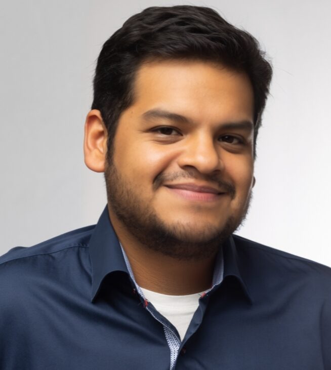
 Jian Ren received his Ph.D. in Electrical Engineering from the California Institute of Technology. After serving as the Chief Technology Officer of oProbeLLC, a start-up company commercializing his graduate work on emerging endoscopy technologies, he performed his post-doc research at Massachusetts General Hospital (MGH). In 2022, he joined the faculty at the Gordon Center for Medical Imaging at MGH and Harvard Medical School. His early work contributed to developments in nonlinear optics and optical communications, including establishing a world record of optical delay using a parametric process. Dr. Ren’s research interests include biophotonics, neurophotonics, biomedical imaging, and nano/micro photonics. He received the Pathway to Independence Award (K99/R00) from the National Institutes of Health.
David Wong-Campos was born in Peru and attended the Monterrey Institute of Technology and Higher Education (ITESM) in Mexico for his undergraduate. He completed his Ph. D. in Physics at the University of Maryland in quantum physics, where he implemented a novel quantum logic gate and performed high-resolution imaging experiments on individual atoms. He took a two-year industry job at a quantum computing startup (IonQ) and held an advisory position at a cancer company (Delee). He returned to academia and currently pursues postdoctoral training with Adam Cohen on voltage imaging of dendrites in vivo.
Zeinab Hajjarian is an Assistant Professor of Biomedical Engineering at the University of Massachusetts, Lowell. She received her Ph.D. in Electrical Engineering from Pennsylvania State University. Her doctoral work focused on modeling light propagation through atmosphere to enable optical telecommunications and imaging. She transitioned to biomedical optics in 2010 by pursuing post-doctoral training at the Wellman Center for Photomedicine, Massachusetts General Hospital. There, she developed technologies for characterizing the mechanobiology of human disease, including cardiovascular disease, coagulation disorders, and carcinoma. Her current research involves development and translation of novel optical microscopes for mapping the biophysical and biomechanical properties of tissues.